
Modeling Intended and Unintended Neurostimulation by EM-fields
Problem Description
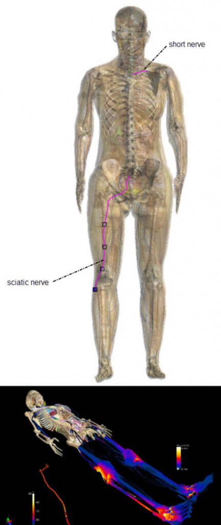
Highly resolved full body human model, with details of the nerves (top); Sim4Life simulation of exposure in an MRI examination.
Electromagnetic fields (EMF) interact with neurons. The interaction can be stimulating, inhibitory, or synchronizing, and it can be intended or unintended. Unintended stimulation by exposure to strong low frequency fields is for example occurring in magnetic resonance imaging (MRI) gradient coils, while examples of intended stimulation include therapeutic applications (transcranial stimulation, deep brain stimulation, functional electrical stimulation, etc.) or neuroprosthetic devices (artificial retina, neuroprosthetic limbs, etc.). Modeling is particularly valuable for treatment and device safety and efficacy assessment, but also to optimize medical device performance.
The prediction of safety thresholds, stimulation selectivity, spiking frequency, pulse-shape impact, etc. is complicated by the intricate structure and ion channel dynamics of the neurons, by the inhomogeneous nature of the electric field distribution in the human body, and by the complex interplay between the two. The latter is the reason, why coupled EM-neuronal dynamics modeling is required.
Related Standards
There are multiple relevant standards regulating EM exposure safety with regard to induced neuronal dynamics: The ICNIRP 2010 exposure guidelines and the IEEE C95.1 exposure standard provide thresholds for general public and occupational exposure to low frequency fields that are based on considerations dominated by the need to prevent adverse EM-neuron interaction-related effects. The IEC 60601-2-33 standard regulates specifically exposure the MRI-related fields.
An important factor in deriving safety limits for the guidelines and standards is the SENN (Spatially Extended Nonlinear Node) model of neuronal dynamics that was designed to represent myelinated axons (nerve fiber).
Methodology
1. Coupled EM-Neuronal Dynamics Modeling in Sim4Life
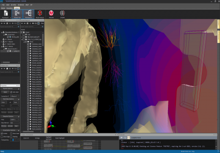
Detail of a simulation of a spinal neural stimulator, in Sim4Life.
The Sim4Life T-NEURO module offers comprehensive neuronal dynamics simulation, full integration and coupling to the EM modeling functionality (P-EM-FDTD and P-EM-QS) of the Sim4Life platform, as well as a range of predefined neuronal dynamics models, including the SENN model underlying the safety standards. A principal strength of Sim4Life is its ability to simulate complex neuronal dynamics models within realistic anatomical models (e.g., the Virtual Population (ViP) 3.0 or models generated from medical image data using the IMG and the iSEG modules). The T-NEURO module is powered by the NEURON solver developed at the Yale University.
Neuron models can be easily designed either by specifying trajectories as splines and then attributing them predefined behavior models, or by importing detailed neuron models from large repositories, such as the ModelDB. Functionality to automatically determine stimulation thresholds for given pulse shapes is available.
2. Application to Neuroprosthetics
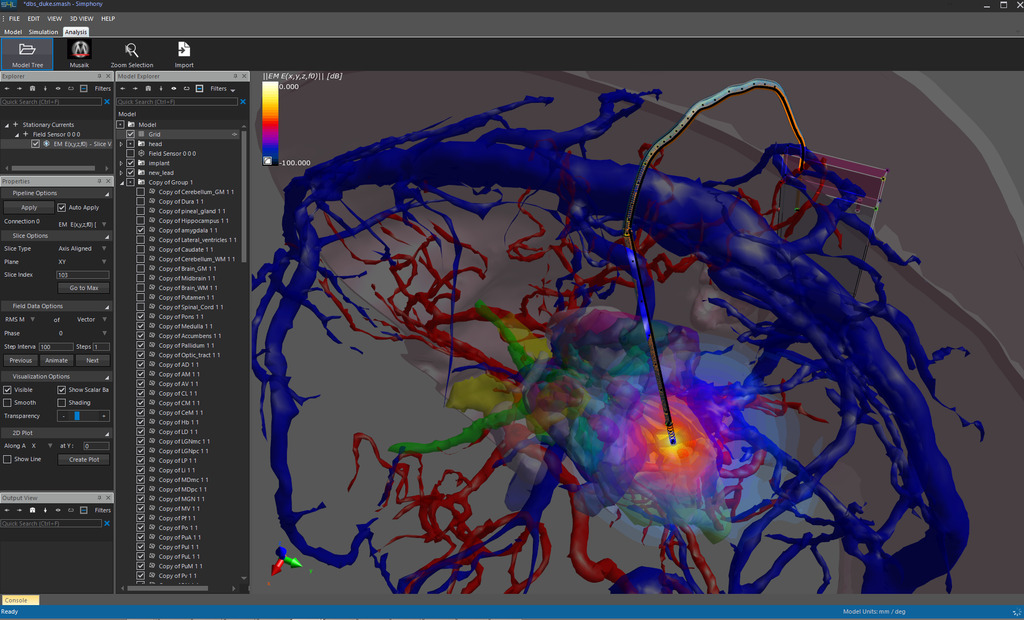
Detail of a simulation of a deep brain stimulator (DBS) in Sim4Life.
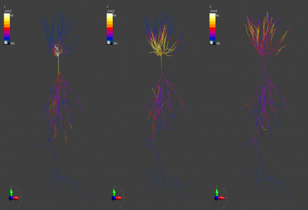
Using the T-NEURO functionality of Sim4Life it is possible to investigate implantable electrodes for neuroprosthetic applications. For example, a transverse intrafascicular multichannel electrode (TIME – a neural interface that promises higher stimulation selectivity at the cost of increased invasiveness when compared, e.g., to more common cuff electrodes) design featuring five sub-electrodes to selectively stimulate different neuron groups in the sciatic nerve related to the activation of various muscles was simulated. For this, the nerve geometry, including the different fascicles was extracted from image data and transformed into a nerve model. Several hundred dynamic neuron models capturing the statistical variability of neuron properties were then placed inside the nerve model and their stimulation by the TIME electrode array modeled. Such simulations were used to compare different electrode designs with regard to muscle stimulation selectivity and placement sensitivity. The simulation predictions were confirmed by experimental measurements of muscle stimulation in rats. Sciatic nerve stimulation with TIME electrodes is being investigated with the goal of returning leg motion to paraplegic and first results in mice are very encouraging.
3. Application to Neurostimulation
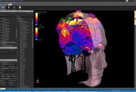
Currents induced by external transcranial magnetic stimulation. Simulation with Sim4Life.
Neurostimulation using external or internal electrodes is applied for various purposes. For example, deep brain stimulation (DBS) uses implanted electrodes to treat movement disorders, depression, etc. Transcranial stimulation uses external electrodes mounted on the head surface, e.g., for stroke rehabilitation. With Sim4Life, it is possible to simulate not only the electric field distribution and currents, but also the related impact on neuronal activity. Field distributions from various transcranial stimulation electrode montages have been compared using the Sim4Life low frequency solvers in combination with the highly resolution MIDA head model and the obtained current densities through the retina could be correlated with the experimentally observed occurrence of visual phosphenes, i.e., the phenomenon of seeing light in absence of light entering the eye.
4. Application to MRI safety
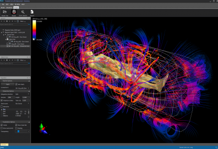
Highly resolved full-body human model, undergoing an MRI examination. Simulation with Sim4Life.
Coupled EM-neuronal dynamics modeling has been applied to assess safety concerns about unintended nerve stimulation induced by MRI gradient coil switching. By integrating realistic motor neuron models along various nerve trajectories inside the human body and investigating stimulation thresholds with regard to the fields induced by a realistic gradient coil model inside the functionalized ViP 3.0 phantom, it could be demonstrated that a series of assumptions underlying current safety standards is problematic. Most importantly, it was found that i) in addition to field strength, field inhomogeneity – as present inside the human body – can be a relevant source of neurostimulation, ii) the SENN model is not always conservative, and iii) the impact of temperature on neuronal dynamics is important, such that coupled EM-neuronal dynamics modeling inside realistic anatomical models is required to properly understand low frequency exposure safety and derive suitable safety criteria. The modeling fidelity is further increased by using inhomogeneous, anisotropic conductivity maps obtained with diffusion tensor imaging. EM modeling of DBS electrode exposure of various treatment relevant thalamic and subthalamic nuclei has been combined with simulations of >100 realistic neuron models representing three different neuron populations (accurately placed using Sim4Life’s Python scripting functionality) and the predicted stimulation rates could be related to experimentally determined quantities.
5. Validation
Simulation of a neuron firing due to electrical stimulation. Simulation with the T-NEURO module in Sim4Life.
The underlying EM solvers have been extensively verified, e.g., using the method of manufactured solutions.
The coupled EM-neuronal dynamics modeling has been verified and validated on multiple levels: The correctness of the Sim4Life implementation was verified by reproducing neuron models from the ModelDB in Sim4Life and by comparing against threshold values obtained using the reference SENN model implementation available from the FDA website for a large variety of pulse durations and shapes. Experimental validation was performed by predicting and measuring i) stimulation thresholds for retinal ganglion cells and ii) muscle activation selectivity from neuroprosthetic sciatic nerve stimulation. Furthermore, qualitative validation of deep brain stimulation modeling was performed against literature data.
Publications
- Reilly, J. Patrick, and Alan M. Diamant. Electrostimulation: theory, applications, and computational model. Artech House, 2011.
- Neufeld, Esra, et al. “Simulation platform for coupled modeling of EM-induced neuronal dynamics and functionalized anatomical models.” Neural Engineering (NER), 2015 7th International IEEE/EMBS Conference on. IEEE, 2015.
- Iacono, Maria Ida, et al. “MIDA: A Multimodal Imaging-Based Detailed Anatomical Model of the Human Head and Neck.” PloS one 10.4 (2015).
- Neufeld, Esra, Ioannis V. Oikonomidis, and Niels Kuster. “Thresholds for interference with neuronal activity.” Electromagnetic Compatibility (APEMC), 2015 Asia-Pacific Symposium on. IEEE, 2015.
- Neufeld, Esra, et al. “Computational platform combining detailed and precise functionalized anatomical phantoms with EM-Neuron interaction modeling.”General Assembly and Scientific Symposium (URSI GASS), 2014 XXXIth URSI. IEEE, 2014.