
Modeling Vagus Nerve Stimulation
Problem Description
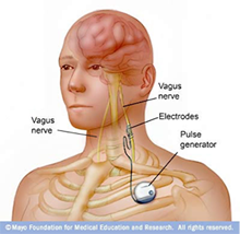
Vagus nerve stimulation (VNS). Image from: http://heatherdane.com/
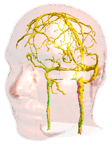
Computational MIDA head model [2], specifically realized for neurostimulation investigations.
Vagus nerve stimulation (VNS) was approved in 1997 by the US Food and Drug Administration (FDA) [1] as an invasive neuromodulator approach for the treatment of epilepsy in anti-epileptic drug (AED) resistant subjects. As the vagus nerve (VN) innervates many organs, it is candidate for many new potentially therapeutically relevant applications of selective neurostimulation.
The VN (like many other large nerves in the human body) is composed by multiple functional units assembling many myelinated A- and B- axons of different diameters combined with small unmyelinated C-fibers. Hereafter, VNS approaches for therapeutically relevant applications require approaches providing high fiber selectivity. Electrode arrays with optimized stimulation waveforms can be used to provide, in theory, such selectivity. Computational models that feature simplified or realistic VN models embedded in realistic human anatomical models along realistic trajectories, together with electrophysiological models of axonal fibers capturing the electric-neuronal interaction, are fundamental for the computationally assisted formulation of new VNS protocols, the design of electrode arrays, the optimization of stimulation waveforms, and the prediction of setup-specific axonal fiber recruitment.
Functionalized VN models, featuring head models, nerve trajectories with an arbitrary numbers of electrophysiological axonal models, can now be created in Sim4Life with the recently added T-NEURO feature in combination with electromagnetic (EM) simulations in the low-frequency range.
Methodology
Sim4Life, with its expanded simulative abilities of T-NEURO, permits the investigation of the mechanisms of interaction between EM fields and the electrical activity of neuronal membranes. A wide range of applications ranging from neurostimulation investigations (e.g. transcranial electric (TES) and magnetic (TMS) stimulation), safety (e.g. against peripheral nerve stimulation (PNS) in magnetic resonance imaging (MRI)), as well as the computationally assisted development of electroceuticals or neuroprostetic devices, is now possible. Anatomically realistic compartmentalized electrophysiological representations of axons and neurons modeled using NEURON [3] libraries according to Sim4Life pre-defined biophysical models or available in the web-depository databases – can be positioned within the computational human head/body models to predict the physiological response of nerve and neurons to applied electric (E-) or magnetic (M-) fields. Highly customizable solutions for modeling, execution of simulations, and post-processing analysis for numerical optimization are also provided in Sim4Life.
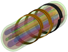
1. Modeling of VN Geometry, Electrodes, and EM Simulations
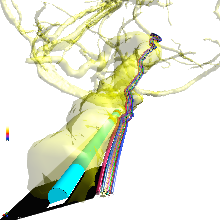
2D section of Helmers’s VN model [4] (top); simplified 3D model (middle); anatomically realistic VN model that follows the trajectory of the VN in the MIDA model [2,5] (bottom).
Cross-sectional 2D VN models that feature, e.g., epineurium, perineurium, and fascicles, can be created from medical images or scratched in Sim4Life (see Figure top) with the graphical user interface (GUI). 3D models of the VN can be created, for example, by extruding realistic 2D nerve cross sections along user-defined trajectories (see Figure middle), or along anatomical nerve trajectories within computational human models (see Figure bottom) Electrode geometries can be imported in Sim4life as CAD files or created as parameterized model objects even using available templates entities (helical, spiral, etc.).
Sim4Life’s electro-quasi-static current dominated (EQSCD) solver can be used to set up low frequency EM simulations for either homogeneous or anisotropic dielectric tissue parameters. Electrode voltages may be assigned as Dirichlet boundary conditions.
Sim4Life’s post-processing tools permit the analysis and visualization of exposure-related quantities such as E-field distribution, input currents at electrodes as well as neurostimulation-related quantities as Eigenvalues and Eigenvectors of the E-field Jacobian to identify regions of neurostimulation on the basis of the ‘activation function concept’ [6].
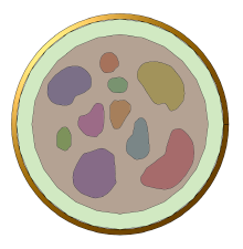
2. Creation of Axonal Trajectories and Functionalization
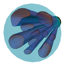
Fascicles populated with axonal trajectories modeled as lines.
具有神经外膜、神经外膜和束等特征的横截面 2D VN 模型可以从医学图像创建或在 Sim4Life 中使用图形用户界面 (GUI) 进行划痕(参见图顶部)。可以创建 VN 的 3D 模型,例如,通过沿用户定义的轨迹(参见图中间)或沿着计算人体模型中的解剖神经轨迹(参见图底部)挤出逼真的 2D 神经横截面,可以将电极几何形状导入 Sim4life 作为 CAD 文件或创建为参数化模型对象,甚至使用可用的模板实体(螺旋、螺旋等)。
Sim4Life 的电准静态电流主导 (EQSCD) 求解器可用于为均匀或各向异性电介质组织参数设置低频 EM 模拟。电极电压可以指定为狄利克雷边界条件。
Sim4Life 的后处理工具允许分析和可视化与暴露相关的量,例如电场分布、电极上的输入电流以及神经刺激相关量,如电场雅可比行列式的特征值和特征向量,以识别神经刺激区域 “激活函数概念”的基础[6]。
3. Execution of T-NEURO Simulation
T-Neuro simulations can be executed serially or in parallel. Each simulation can include multiple independent electric sources of neurostimulation (as in the case of electrode arrays), each with its own stimulation waveform.
Point or line sensors can be defined to record transmembrane potential or currents information, to be used for post-pro analyses. The implemented ‘titration procedure’ is fundamental to identify threshold values of E-field or stimulation related quantities (such as input currents) for initiation of action potentials (APs) within each individual axon in the model.
4. Post-processing
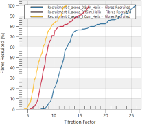
Visualization of the activation function along fiber trajectories (top); examples of fiber recruitment curves for three types of axons populations (bottom).
Sim4Life provides multiple options for data visualization (slice and surface view, vector field view and streamline, etc.) including the calculation of E-field related integrals (i.e., flux integrator), the visualization or animation of transmembrane voltages or current profiles. These features can be also used for the visualization of customized post-pro quantities derived from python script (e.g., compound action potentials).
The activation function, predictor of the site of neurostimulation, can be visualized along the axonal geometries (see Figure top). The titration sensor provides threshold E-fields for spike initiation, time and location of spike initiation and permits to derive fiber recruitment curves (Figure bottom).
All post-processing results can be exported for further analysis in MATLAB or Excel. Python scripts and the Sweeper tool can be used to customize optimization procedures aimed, for example, at identifying steering parameters for selective stimulation (e.g. recruitment of either A-, B-, or C- fibers) or to optimize electrodes geometry.
结论
Sim4Life T-Neuro is conceived to assist in the computational assisted investigation of neurostimulation, the design and optimization of electroceuticals and neurostimulators, and for studies about the mechanisms of electromagnetic-neuronal interaction. In this example the benefits of using T-Neuro to investigate fiber recruitment on realistic or simplified models of Vagus Nerve have been illustrated. Similar procedures can be applied to model arbitrary complex nerve stimulation in arbitrary computational human or animal bodies. Sim4Life provides unique and unprecedented features to investigate the complex domain of neurostimulation, affirming itself as the leader tool in the market for neurostimulation investigations